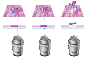
 Genome, transcriptome and proteome analyses require pure samples to obtain reliable results. However, tissue preparations are usually inhomogeneous and consist of a mixture of different cells. As tissue complexity can affect the outcome and interpretation of molecular studies, isolation of pure cell populations is preferred. Using a laser coupled to a research microscope, defined target cells or even subcellular components like organelles and chromosomes, can be isolated. In our lab, a P.A.L.M. microbeam from Zeiss, an inverted microscope with a pulsed UVa-laser, is present.
Advantages of laser capture microdissection
- Obtaining pure cell populations without the risk of contamination
- Genome, transcriptome and proteome analysis after isolation
- Can be performed on different sample types such as snap frozen or formalin fixed paraffin-embedded tissue sections, cytospins, cell smears and chromosome preparations.
- Can be combined with fluorescence staining to visualize particular cells or cell organelles of interest
Workflow
You provide us a glass slide with the cells of interest, stained if necessary to visualize the appropriate cell types. These cells will be isolated directly in a DNA or RNA extraction buffer, upon which DNA or RNA analysis can be performed.