 Flow cytometry is an ideal technique to count cells and to monitor a (live) cell population simultaneously for multiple parameters, such as size (distribution) and morphological complexity. Additionally, a wide range of fluorophores can be used to stain different cell surface and/or intracellular markers, and to label certain cell types making relative quantification of a particular cell type possible. It can also be used for apoptosis assays, for determining cell proliferation and to follow the distribution of the population among the different cell cycle phases.
Advantages of flow cytometry
- Fast and highly informative analysis
- Simultaneously analyzing multiple cell markers
- High-throughput sample analysis possible
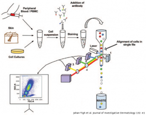
Workflow
Depending on the analysis of interest, we start from a live cell sample directly from cell culture or cells isolated from a biological sample such as blood. Those samples can be run immediately, if only cell count, size, and morphology need to be determined. If also monitoring one or multiple biomarkers (e.g. CD-markers) is required, an additional staining step will be included prior to analysis, to label the cells of interest with fluorochrome-coupled antibodies. Likewise, for apoptosis assays, a specific staining protocol will be implemented.
To assess a cell line’s proliferation rate, a specific fluorescent dye (e.g. CFSE) is added to the culture medium after which the cells can be kept in culture for as long as requested. Following this additional incubation step and collecting the samples, the successive cell generations are quantified.
For specific cell cycle experiments, the cells will be permeabilized after harvesting and stained with the appropriate DNA-binding dye (e.g. propidium iodide).
All experiments are performed with a FC500 cytometer (Beckman Coulter) combined with the according CXP analysis software. Five colours can be detected: FITC, PE, ECD, PE-Cy5 and PE-Cy7.